DIAGNOSIS OF
GLAUCOMA

STEP 1

A regular comprehensive Eye Examination including checking of eye pressure for symptoms of Glaucoma. Plus checking for any signs of “cupping” in the optic disc.

STEP 12

If the fluid eye pressure(67. IOP) is elevated, further examinations will be needed and referral to the VE Medical ophthalmologist.

FURTHUR EXAMINATIONS

These additional examinations are needed to fully quantify any loss of vision and the extent of damage to the optic nerve.

VISUAL FIELD TEST

The visual fields test is to check if any peripheral vision has been lost. This is normally the first “vision loss”  as the glaucoma disease progresses

VE Medical have two different options to test peripheral vision.

The HUMPHREY Visual Field equipment and the more recent Headset imaging HERU equipment.
HUMPHREY Equipment
HERU Equipment

With either test,  you are asked to concentrate, focus straight ahead, and flashing lights appear around the edges of your vision. You press a button, every time you see a flashing light. This is a painless test, and takes a lot of concentration, but the VE MEDICAL nurse will guide you through it. The printed results from the visual field test will show the ophthalmologist if you have lost any peripheral vision.

FURTHER DIAGNOSTIC TESTS BY THE DOCTOR
  • Pachymetry … which measures the thickness of the Cornea 
  • Gionoscopy ….A special lens is used to see the outflow of the aqueous liquid in your eye,  and determine if there are any blockages, which are causing the build up of fluid pressure.
  • Tonometry … A further check on the eye pressure as a “double check” 
Screen Shot 2022-10-17 at 8.50.19 AM
Pachymetry
lens
Gionoscopy
pressure-check
Tonometry

THE MAIN FOCUS OF ALL GLAUCOMA TREATMENT IS TO DELAY DAMAGE TO THE OPTIC NERVES

The optic nerves relay messages from your eyes to your brain to create visual images. They play a crucial role in your ability to see. Millions of nerve fibers make up each optic nerve. Damage to an optic nerve can lead to vision loss in one or both eyes.

DIAGNOSIS OF DAMAGE TO THE OPTIC NERVE 

In the last thirty years , the development of wavefront technology has made it possible to do a full examination of the optic nerve. This is called OPTICAL COHERENCE TOMOGRAPHY(OCT) Optical coherence tomography (OCT) is a noninvasive diagnostic imaging procedure  that enables ophthalmologists to understand  different layers of the optic nerve and retinal nerve fiber layer (RNFL) with astounding detail. OCT is an  excellent  tool for screening, diagnosing, and tracking the progression of glaucoma in patients. VE Medical Team use one of the latest Zeiss Cirrus OCT machines to obtain  accurate diagnosis
OCT - OPTICAL COHERENCE TOMOGRAPHY

MORE INFORMATION ON THE SERVICES AT VISION EXPRESS MEDICAL

ORANGE GROVE PLAZA ST. LUCIA

INFO@VISIONEXPRESSCARIBBEAN.COM

Testimonials

"I got my cataract surgery at VE Medical in Saint Lucia. The team took great care of me from start to finish. Very lovely people."
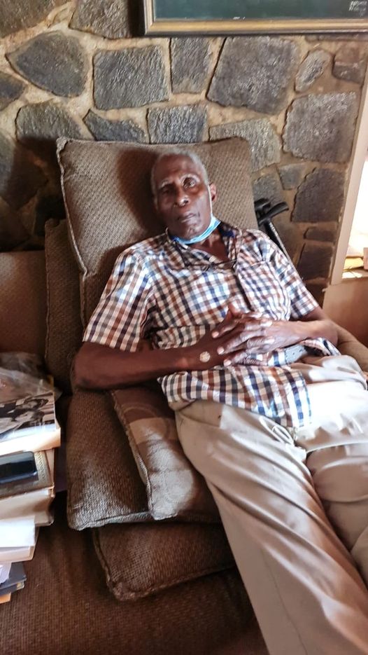
Mr. Fics
Business Man
Ms Caroline Casimir was successfully with cataract and retinal surgery in both eyes."The team at Vision Express are very caring and capable of doing what they do."
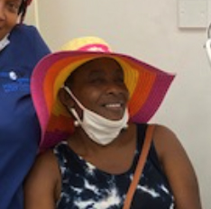
Ms. Caroline Casimir
Ms Dorothy Henry has undergone CATARACT SURGERY in both eyes. She was delighted with the service and was very thankful of the team of nurses and doctors.
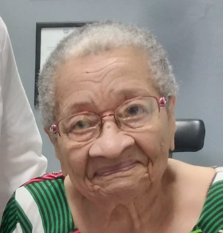
Ms Dorothy Henry

Subscribe to Our Newsletter